020 71832792
 info@privatedoctor.london

Chronic Maxillary Sinus Fullness vs Odontogenic Sinusitis: Navigating Combined Clinical Evaluation
Persistent pressure in the cheeks, upper jaw discomfort, blocked nasal passages, and a dull sense of facial heaviness are common complaints in primary care and dental settings. The diagnostic challenge is that similar symptoms can arise from very different causes. Some patients have chronic rhinosinusitis driven by inflammatory or allergic mechanisms, while others have sinus inflammation secondary to a hidden dental source. Distinguishing these pathways matters because treatment priorities differ, and symptom-only approaches often delay resolution.
This guide explains how chronic maxillary sinus fullness differs from odontogenic sinusitis, how clinicians evaluate overlap cases, which red flags require urgent review, and why coordinated dental and ENT assessment can be decisive in complex presentations.
Introduction: Why This Distinction Is Clinically Important
The maxillary sinuses sit close to the roots of upper premolars and molars. Because of this anatomy, dental pathology can involve the sinus floor, and sinus disease can produce referred discomfort in upper teeth. Patients may therefore circulate between self-treatments for “sinus pressure” and “toothache” without a clear diagnosis.
Misclassification risks include:
- repeated short antibiotic courses without source control
- delayed dental treatment in true odontogenic disease
- delayed ENT intervention in non-dental chronic sinus inflammation
- avoidable chronic symptom burden
A structured differential diagnosis avoids these pitfalls and improves outcomes.
Maxillary Sinus Fullness: Core Concepts
What patients usually mean by “fullness”
“Fullness” typically describes pressure, weight, congestion, or dull ache over one or both cheeks. It may be associated with:
- nasal obstruction
- postnasal drip
- altered smell
- pressure that changes with posture
- facial discomfort during upper respiratory episodes
Common non-dental contributors
- chronic rhinosinusitis (with or without polyps)
- allergic rhinitis with secondary sinus obstruction
- mucosal oedema after viral infection
- anatomical variants affecting drainage pathways
- environmental irritants and occupational exposures
Not all chronic fullness indicates bacterial infection. Inflammatory pathways are frequently dominant and require targeted management rather than reflex antibiotics.
Odontogenic Sinusitis: What Makes It Different
Odontogenic sinusitis refers to maxillary sinus inflammation where the initiating source is dental or dentoalveolar. Typical triggers include periapical infection, periodontal disease, oro-antral communication, and complications of dental procedures.
When evaluating possible dental-source sinus disease, clinicians often review educational references on dental abscess signs and causes because deep endodontic infection can present subtly before overt dental pain appears.
Why laterality matters (but does not diagnose by itself)
Odontogenic disease is often unilateral, but unilateral symptoms are not diagnostic alone. Non-dental unilateral disease also occurs, so imaging and clinical correlation are essential.
Typical clinical clues that increase suspicion
- recent upper posterior dental treatment
- persistent unilateral malodorous discharge
- upper molar/premolar discomfort with sinus symptoms
- prior temporary response to antibiotics with recurrence
- chronic symptoms despite standard rhinosinusitis treatment
Shared Anatomy: Why Teeth and Sinuses Confuse the Picture
The floor of the maxillary sinus may be separated from posterior tooth roots by a thin bony plate, and in some individuals roots project very close to sinus mucosa. This proximity explains:
- referred pain in either direction (tooth-to-sinus, sinus-to-tooth)
- higher sinus vulnerability when upper posterior infections are untreated
- procedural risk windows in extractions, implants, and sinus-adjacent surgery
For implant-related context, clinicians may reference material discussing dental implants near the sinuses, especially where prior surgery complicates interpretation.
Clinical Assessment Pathway: History, Examination, Differential
Focused history
Useful history points include:
- exact timeline and progression
- unilateral vs bilateral symptom dominance
- smell disturbance and discharge quality
- prior dental pain, root canal, extraction, implant placement
- previous response to nasal steroids, saline, or antibiotics
- smoking status and comorbidities
Dental examination components
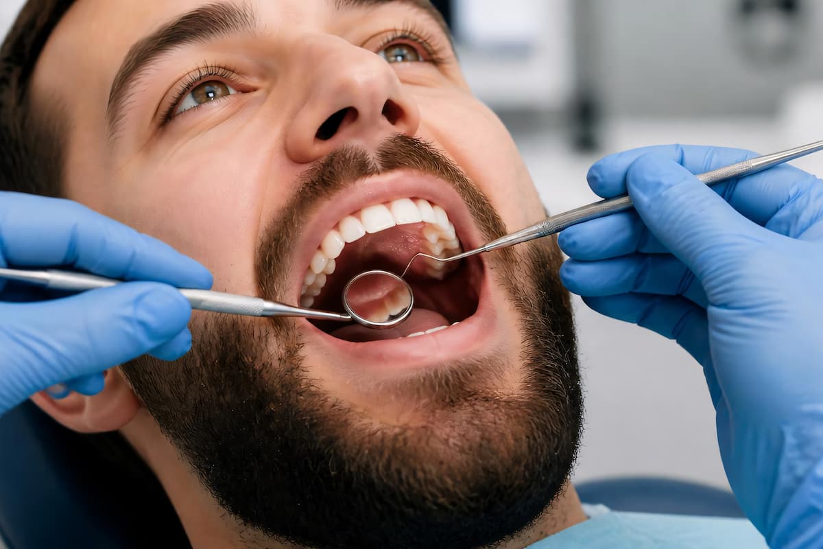
Dental review assesses:
- percussion tenderness
- periodontal pocketing
- tooth vitality
- mobility and occlusal pain
- signs of fistula or gingival swelling
This is where a formal dental examination and assessment services pathway is often valuable for structured source identification.
Periodontal disease as a contributor
Chronic periodontal burden can coexist with endodontic pathology and sustain low-grade inflammatory load near the sinus floor. Educational resources on periodontal disease affecting the gums are often used to explain why gum status matters in sinus-adjacent cases.
ENT examination components
- anterior rhinoscopy
- targeted endoscopy (where indicated)
- assessment of middle meatus drainage patterns
- evaluation for polyps, oedema, and anatomical obstruction
Imaging Strategy: Choosing the Right Tool for the Question
No single test answers every question. Imaging selection depends on clinical suspicion.
Dental radiography and CBCT
Best for:
- periapical pathology
- root morphology
- sinus floor relationship
- focal bony changes
Sinus CT
Best for:
- mucosal thickening patterns
- ostiomeatal complex involvement
- wider sinonasal disease burden
- pre-procedural mapping in advanced cases
Correlation principle
Radiology findings must be interpreted with symptoms and exam. Incidental mucosal thickening alone does not prove the primary symptom driver.
Management Logic: Treat the Source, Not Just the Symptoms
Non-dental dominant pathway
When sinus disease is primarily inflammatory/non-dental, management may include:
- saline irrigation
- intranasal corticosteroids
- trigger control (allergic/environmental)
- guideline-based ENT follow-up
Dental-source dominant pathway
When odontogenic origin is likely, source control is central:
- endodontic treatment where tooth salvage is appropriate
- extraction for non-restorable or high-risk teeth
- periodontal treatment where supporting structures are involved
- adjunctive ENT measures if sinonasal inflammation persists
In selected root-source cases, the care pathway may include root canal treatment for infection where clinically suitable.
Antibiotics: Useful in Context, Inadequate Alone
Antibiotics may be appropriate when acute bacterial signs are present, but repeated empiric courses without source control can mask symptoms while disease persists. This is especially true when dental pathology remains untreated.
Principles include:
- culture/imaging-informed escalation in refractory cases
- shortest effective course consistent with guidelines
- source-directed intervention rather than serial empiric prescribing
Why Combined Dental–ENT Care Improves Outcomes
Complex overlap cases benefit from parallel reasoning:
- dental team defines tooth and periodontal source risk
- ENT team defines sinonasal drainage and mucosal burden
- integrated plan sequences interventions to reduce recurrence
Without this coordination, patients may receive partial treatment in the wrong order, prolonging symptoms.
Practical Symptom Patterns and What They Suggest
Pattern A: Bilateral pressure with allergic history
Often points toward inflammatory rhinosinusitis rather than odontogenic disease, though dental co-pathology is still possible.
Pattern B: Persistent unilateral symptoms with foul smell
Raises suspicion of odontogenic contribution, especially with recent upper dental intervention.
Pattern C: “Sinus pain” plus bite tenderness in one upper quadrant
Suggests combined evaluation for endodontic and sinus disease.
Pattern D: Recurrent “sinus infections” despite multiple antibiotics
Requires reassessment of source diagnosis and imaging strategy; resistant patterns often indicate unresolved local pathology rather than simple recurrent viral illness.
High-Risk Features Requiring Prompt Escalation
Urgent clinical review is needed for:
- periorbital swelling or visual changes
- severe unilateral facial pain with systemic illness
- high fever with neurological symptoms
- rapidly progressive swelling
- trismus or deep neck symptom progression
Emergency pathways are required if orbital, intracranial, or deep-space complications are suspected.
Post-Treatment Follow-Up: Preventing Chronic Recurrence
Resolution is judged by both symptom improvement and objective reassessment where indicated.
Follow-up should review:
- persistence of unilateral symptoms
- residual foul discharge
- dental pain on function
- need for repeat imaging in non-resolving cases
- preventive oral and nasal care habits
Recurrence reduction strategies include:
- regular dental maintenance and periodontal surveillance
- early management of upper posterior dental symptoms
- control of allergic and inflammatory nasal factors
- timely reassessment if symptoms relapse
Communication Tips for Patients Attending Appointments
Patients often improve diagnosis speed by bringing:
- a timeline of symptom onset and progression
- details of all prior dental work (especially upper molars/premolars)
- previous imaging reports if available
- list of completed medicines and response quality
- specific triggers (posture, temperature, exercise, meals)
Clear chronology frequently reveals source patterns that isolated snapshots miss.
Recovery Timelines and Expected Clinical Milestones
Patients often ask how quickly they should improve once a treatment plan starts. Timelines vary by cause and chronicity, but setting expectations helps avoid premature treatment switching.
Early phase (first 1-2 weeks)
- reduced pressure intensity may occur before complete symptom resolution
- nasal discharge quality may improve before volume decreases
- dental-source pain may settle quickly after source control, while sinus discomfort lags
Intermediate phase (2-8 weeks)
- mucosal inflammation can continue to settle after dental intervention
- smell disturbance may recover gradually rather than suddenly
- intermittent pressure fluctuations may occur, especially during colds or allergen exposure
Longer phase (2-3 months and beyond)
- persistent unilateral symptoms despite source treatment need re-evaluation
- recurrences often indicate either incomplete source control or coexisting inflammatory disease
- objective review (repeat examination and, where needed, imaging) is preferable to repeated empiric medication changes
Patients should be advised that partial early improvement is common and does not necessarily mean treatment failure. The key issue is directional trend: stable improvement versus relapse pattern.
Practical Prevention Strategy for Recurrence Reduction
Because many cases are multifactorial, recurrence prevention should combine dental, nasal, and behavioural measures rather than relying on a single intervention.
Dental domain
- maintain regular periodontal and restorative follow-up in upper posterior segments
- seek prompt review for new bite pain, swelling, or prolonged sensitivity
- avoid delaying treatment of suspected periapical disease
Sinonasal domain
- continue clinician-recommended nasal maintenance where inflammatory disease coexists
- monitor seasonal triggers and environmental irritants
- reassess persistent obstruction rather than repeatedly self-medicating
System-level habits
- keep a concise record of symptom laterality and trigger context
- complete planned follow-up rather than stopping at partial symptom relief
- return early if unilateral foul discharge recurs
In practice, this combined approach is what most consistently prevents the cycle of temporary improvement followed by recurrent pressure and repeated antibiotics.
Frequently Asked Questions
Can sinus fullness occur without infection?
Yes. Inflammation, drainage dysfunction, and allergic factors can cause persistent fullness without bacterial infection.
Does unilateral sinus pressure always mean a dental cause?
No. It increases suspicion but does not confirm diagnosis. Examination and imaging are needed.
Can a tooth problem exist with minimal tooth pain?
Yes. Some endodontic or periodontal pathology causes subtle symptoms but still affects adjacent sinus tissue.
Should antibiotics be started before dental review?
Only under clinician guidance. In many recurrent cases, source control is more important than repeated empiric antibiotic cycles.
Are scans always necessary?
Not always. Imaging is selected based on clinical probability and intended management decisions.
Conclusion
Chronic maxillary sinus fullness and odontogenic sinusitis can appear similar but require different treatment priorities. The key clinical task is identifying the dominant source and sequencing care accordingly. Symptom suppression alone rarely solves recurrent or unilateral disease. A coordinated dental and ENT approach, supported by targeted imaging and source control, provides the most reliable pathway to durable improvement.
Disclaimer
This article is for general educational purposes only and is not a diagnosis or personalised medical advice. Symptoms, risks, and test suitability vary by individual and should be assessed by a qualified clinician. Seek urgent care or call 999 for severe or rapidly worsening symptoms.
Written Date: 26 October 2026 | Next Review Date: 26 October 2027